Unsupervised Leaf Vein Segmentation
W-Net for Plant Phenotyping
Deep Learning - University of Tennessee
W-Net, Unsupervised Segmentation, PyTorch
Unsupervised Image Segmentation for Plant Phenotyping
Training a W-Net to segment leaf veins from over 14,000 leaf scans, without a single hand-labeled pixel.
Authors: Hashan Fernando, Peter Kruse
Additional Resources: Paper (PDF) · Git Repository
Plant phenotyping, measuring and characterizing a plant's expressed traits, is a bottleneck in genetic and breeding research precisely because it's so manual. Leaf vein segmentation is a good example: understanding how veins respond to drought or other treatments requires isolating vein pixels from the rest of the leaf, but the deep learning models that do this well are almost all supervised, and hand-labeling veins at the pixel level across thousands of high-resolution leaf scans is exactly the kind of tedious, expensive process phenotyping is trying to escape.
This project trains a W-Net, an unsupervised segmentation architecture that pairs two U-Nets in an encoder-decoder loop, to segment leaf veins with no labeled training data at all. The encoder produces a pixel-wise segmentation map directly from an RGB leaf image; the decoder reconstructs the original image from that map. Training balances two loss functions: soft normalized cut loss, which pushes the encoder toward coherent segment boundaries, and reconstruction loss, which keeps those segments faithful to the original image. We trained and evaluated it on over 14,000 scanned leaf images from a UC Davis plantation, with preliminary testing on an open Ficus Religiosa ("Pimpal") leaf dataset, and compared results against a state-of-the-art supervised vein-segmentation baseline.
From leaf scan to segmentation map
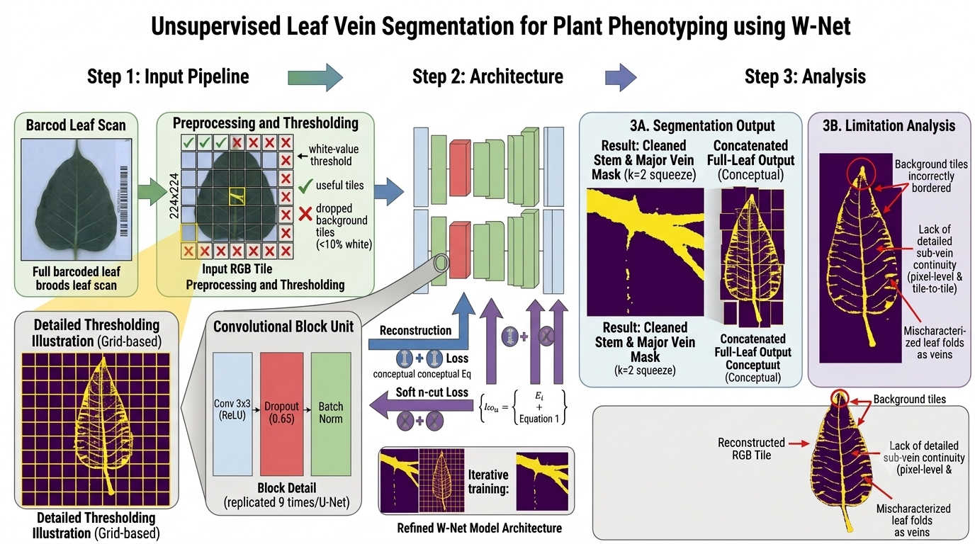
Four stages take a full-resolution leaf scan to a learned vein segmentation, with no labels involved in training.
Tiling & Filtering
Leaf scans (3510×2550 px) are too large to fit in memory, so each is split into 224×224 tiles. Tiles that are more than 10% white background pixels are dropped, keeping the model focused on actual leaf structure.
W-Net Architecture
Two symmetric U-Nets, nine convolutional blocks each, with skip connections between matching contractive and expansive layers, form an encoder (Uenc) that segments and a decoder (Udec) that reconstructs from that segmentation.
Dual-Loss Training
Each batch is optimized in two steps: soft normalized cut loss shapes the encoder's segmentation quality, then reconstruction loss (comparing Udec's output to the original tile) is minimized across the full network; no ground-truth labels required either time.
Postprocessing & Evaluation
Predicted tiles are reassembled by their saved grid index into a full-leaf segmentation map, then compared pixel-wise against eight manually labeled test leaves and against a supervised segmentation baseline.
The full pipeline, end to end
Tools & Stack
What the model actually learned
Tested first on an open leaf dataset, then on 14,000 UC Davis scans, with an eye toward where an unsupervised approach breaks down.
How many segmentation classes best separate veins from leaf body?
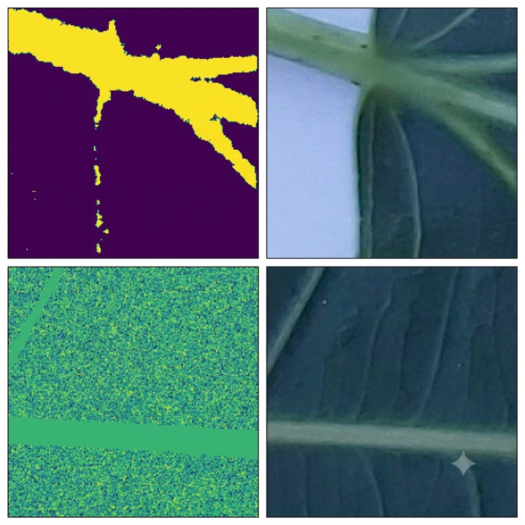
The squeeze parameter k controls how many classes the encoder's output layer predicts. We compared k=2 (vein vs. non-vein) against k=64 on identical leaf tiles.
Solution
k=2 produced the clearest, most interpretable vein segmentations. k=64 could still pick out major veins, but the leaf body and finer veins came out noisy, so every later experiment used k=2.
Does a model trained on 224×224 tiles hold together when reassembled into a full leaf?
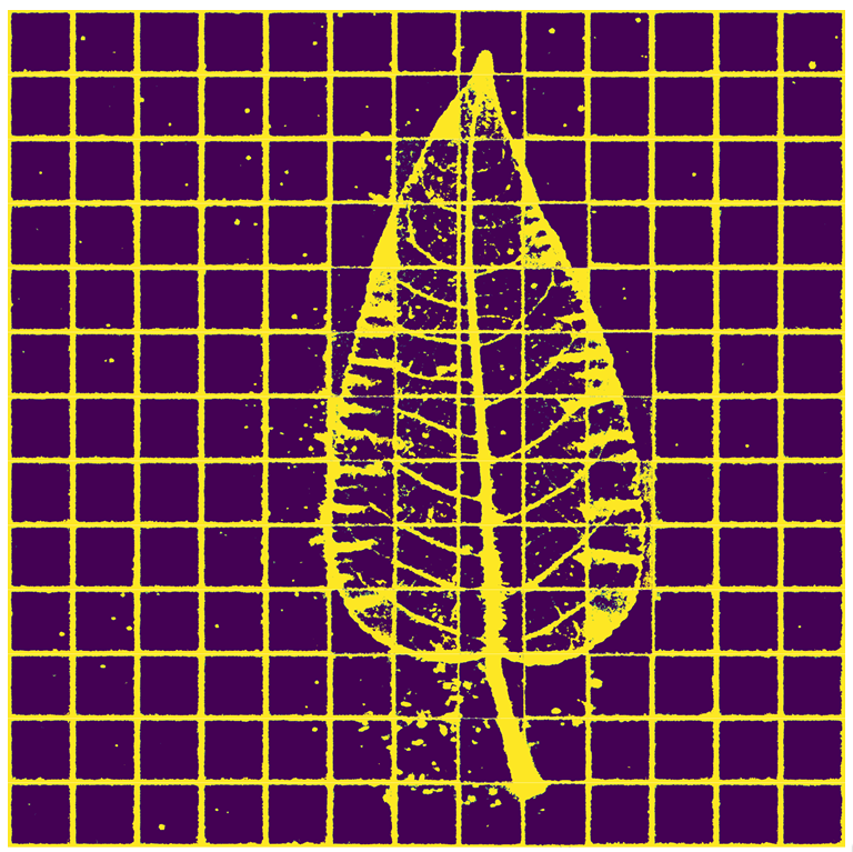
Individual tiles trained well in isolation, but a full leaf image requires hundreds of independently-classified tiles to agree with each other at their shared borders.
Solution
The reassembled maps clearly capture major veins and the leaf-body boundary, but tile-to-tile vein continuity breaks down, blank background tiles pick up spurious borders, and discolored or unhealthy leaf regions get segmented as if they were veins, the model was learning texture contrast in general, not veins specifically.
We were genuinely impressed that W-Net could learn coherent vein structure with zero labeled pixels, but it isn't yet a replacement for supervised, state-of-the-art vein segmentation. The clearest next steps: incorporating principal curvature analysis to enforce vein continuity, switching to a loss function better suited to tiled high-resolution inputs, and testing whether adjusting k further could help the model separate damaged or discolored leaf regions from true veins instead of confusing the two.
Figures